|
||||||||||||||||||
|
||||||||||||||||||
|
|
||
|
 |
 |
 |
 |
 |
|
||||
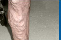
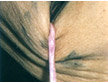
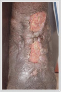
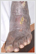
Venous Ulcer
Aetiology
Causes of Venous Ulcers
Pathophysiology of Venous Ulcer
Initial Assessment
Treatment of Venous Ulcer
Dressings Four layer Compression Bandaging
Stemmer et al
All bandages used are 10 cms. in width and should
be applied from the base of the toes to the knee joint.
Layer II
Layer III
Layer IV
Contraindication
|
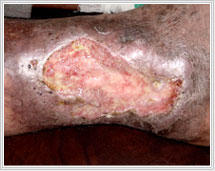 Leg
ulcers are treated by different forms of dressings by the
family physicians. It is extremely important to know what
the cause for the ulcer is? 80% 0f the ulcers in the legs
are due to vascular insufficiency. Healing and preventing
recurrences can only be achieved by correcting the vascular
cause. Dressings do play an important role in aiding ulcer
healing.
Leg
ulcers are treated by different forms of dressings by the
family physicians. It is extremely important to know what
the cause for the ulcer is? 80% 0f the ulcers in the legs
are due to vascular insufficiency. Healing and preventing
recurrences can only be achieved by correcting the vascular
cause. Dressings do play an important role in aiding ulcer
healing.
|
Site Designed and Hosted
by Layout
Galaxy
|